Welcome to our department dedicated to advanced ocular imaging! We are excited to share with you the fascinating world of cutting-edge techniques and imaging technologies used to capture highly detailed images of the eye’s structures and tissues.


Optical coherence tomography (OCT) stands out as one of the most remarkable advancements in ocular imaging. By employing light waves, OCT creates cross-sectional images of the eye, enabling us to detect subtle changes, measure thickness and volume, and diagnose conditions such as keratoconus (KC), macular degeneration, glaucoma, and diabetic retinopathy (DR).


We actively work on developing sophisticated, next-gen tools and automated algorithms for quantitative evaluation of both Corneal and Retinal OCT cross-sectional images.


Cornea

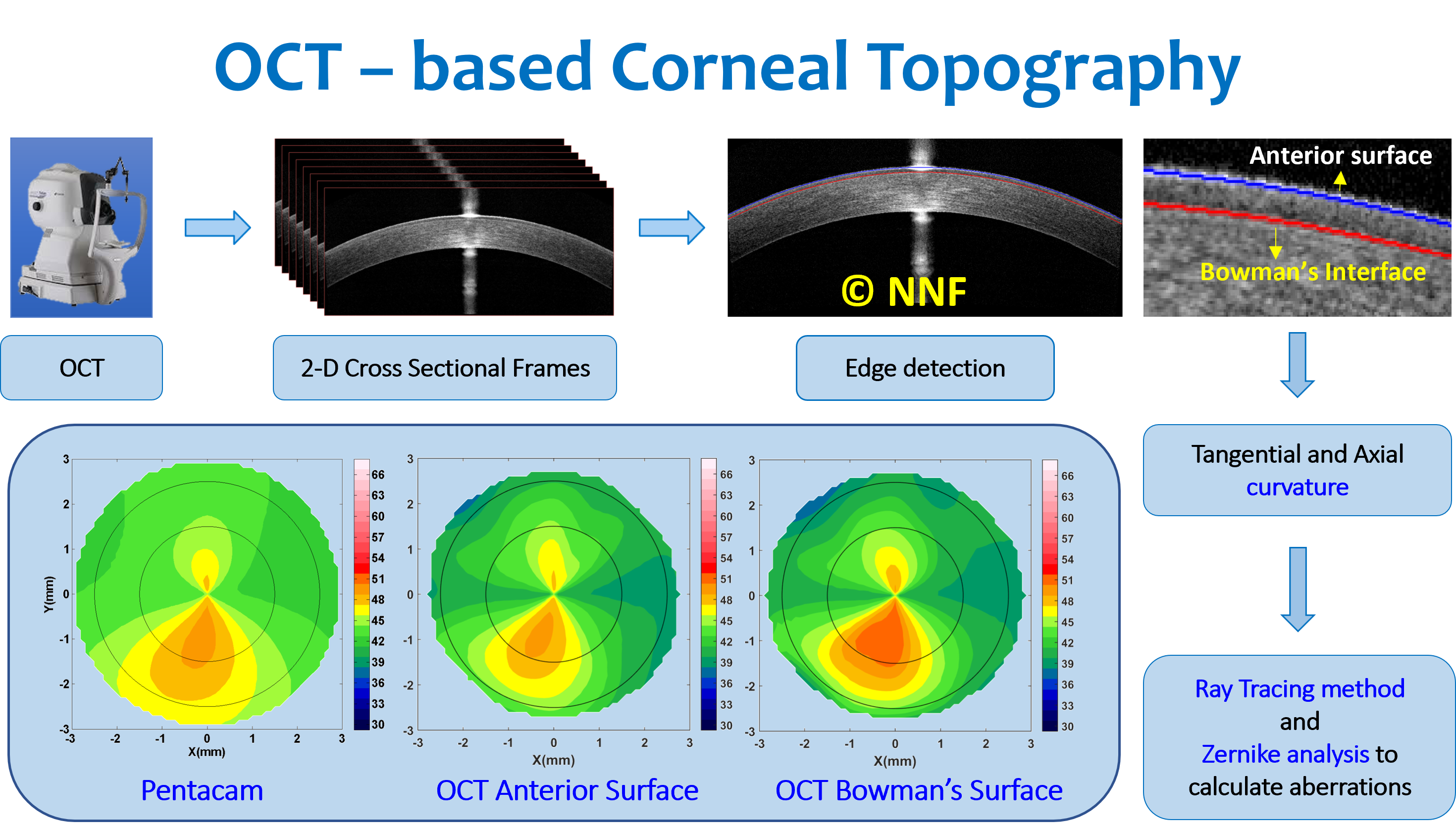
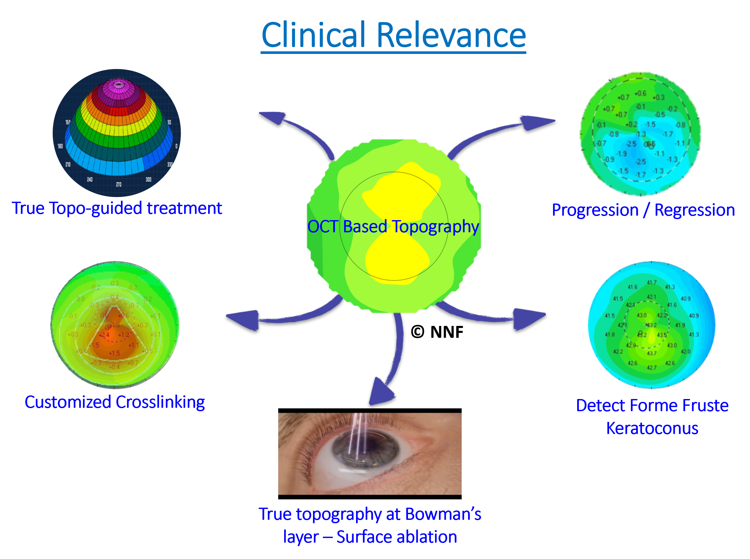
In Cornea, we were one of the firsts in the field to develop anterior corneal and Bowman’s layer topography (curvature) and aberration maps from high-resolution OCT images. This continues to help our clinicians for improved detection of early ectasia.


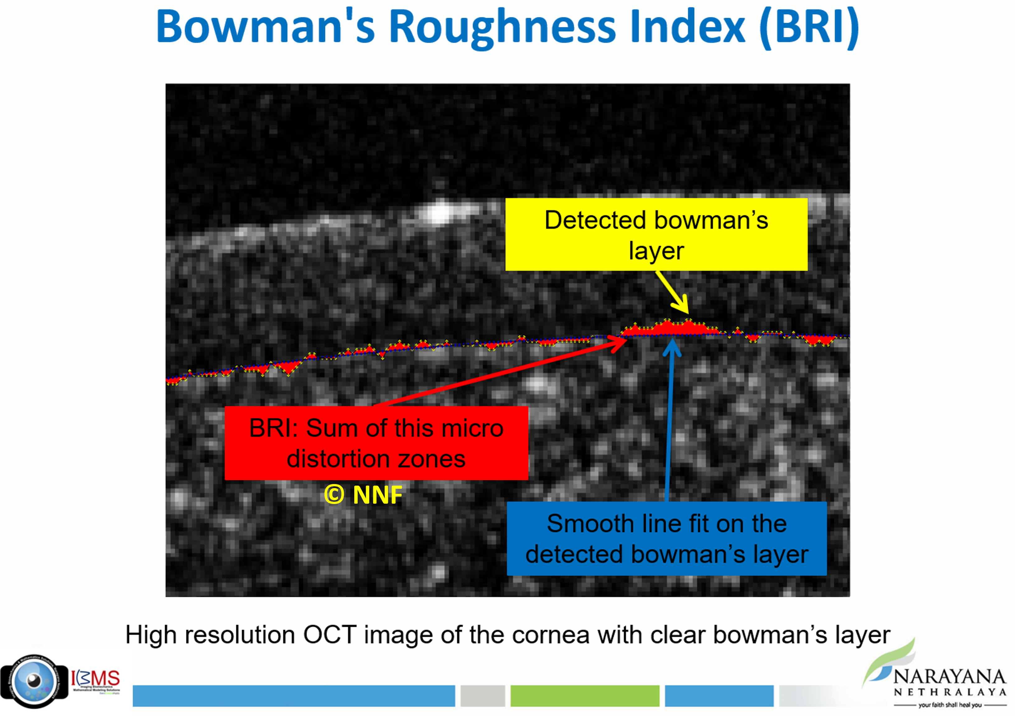
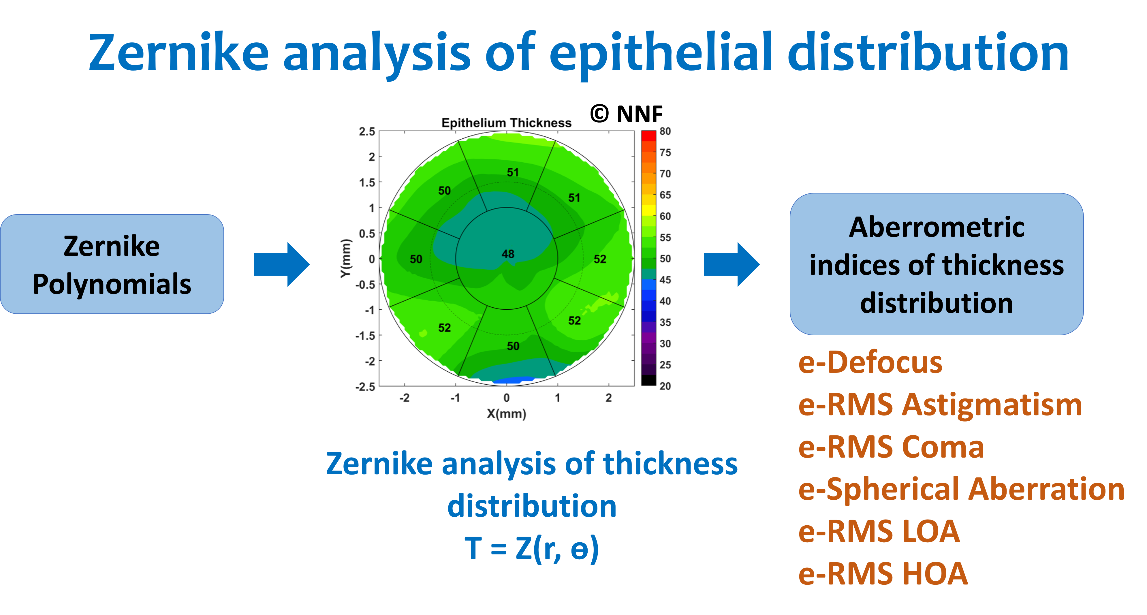
Using high-resolution OCT images, we have serially evaluated the differential remodeling of the epithelial layer after refractive surgeries like LASIK, SMILE, PRK, etc. Extending this further, we have quantified the irregularities and micro-distortions in the Bowman’s layer from high-resolution OCT images and its diagnostic capabilities to help in identifying asymmetric keratoconus.


Our area of research in Cornea also focuses on the effect of different refractive surgeries on patient-specific improvement in intermediate and near accommodation and quality of vision.

Retina

In the Retina, we have developed automated algorithms and tools to quantify high-resolution OCT images of diseases like glaucoma, DR, etc. With spectral domain OCT (SD-OCT) techniques like enhanced depth imaging (EDI), enhanced vitreous imaging (EVI), etc we have compared and evaluated structural visibility of posterior vitreous for routine clinical examinations. 


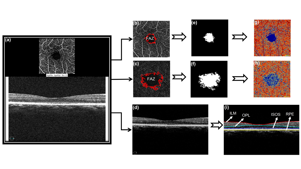
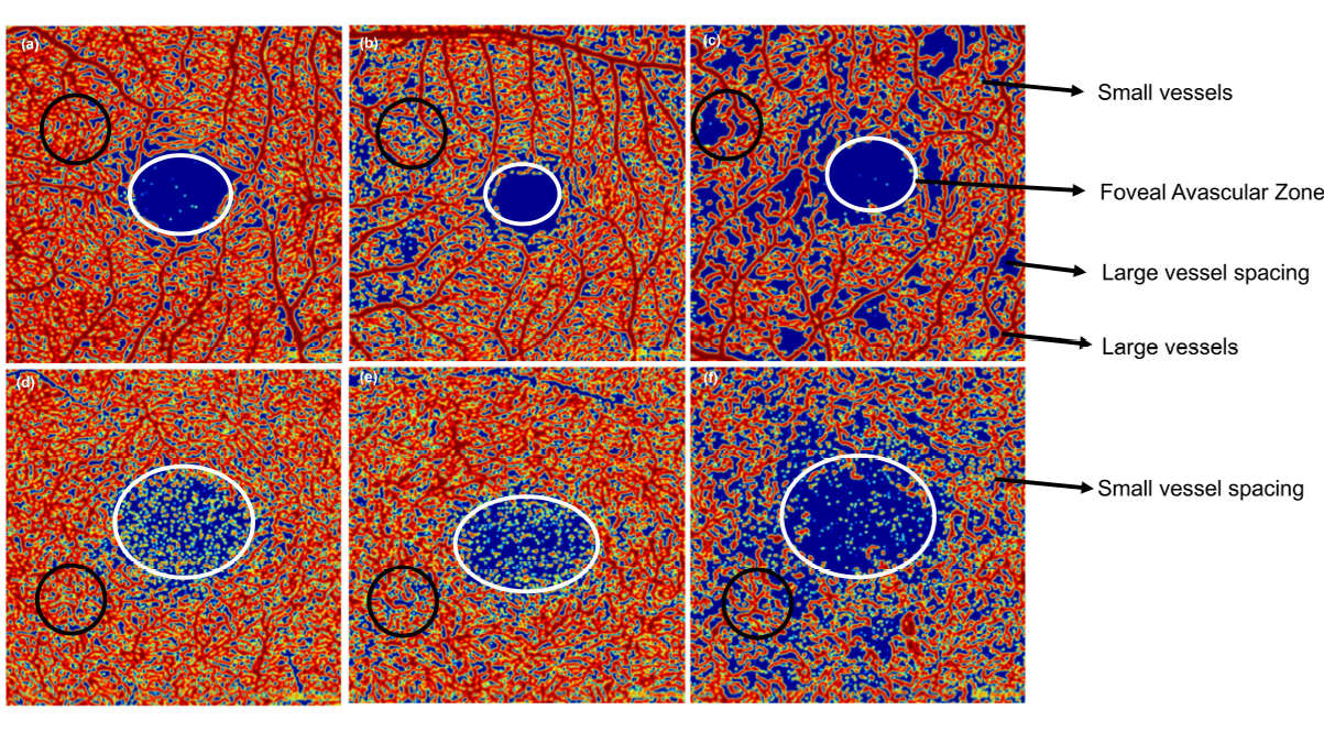
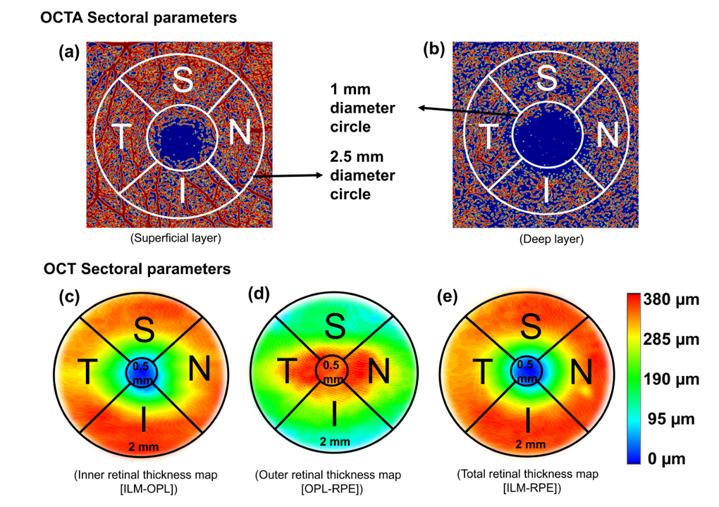
Using OCT Angiography (OCTA), we have extensively compared and quantified retinal vascular features between multiple devices. Our studies have found significant correlation of volume of macular edema with retinal tomography features in patients with DR.

Polarization-Sensitive Optical Coherence Tomography (PS-OCT)


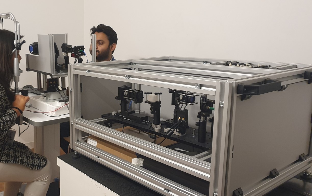
Our most recent addition is the ultra-high-resolution (UHR) Polarization-Sensitive OCT (PS-OCT). This next-generation imaging device was built as a collaborative effort with some brilliant scientists and researchers from the Medical university of Vienna, Austria in 2017.


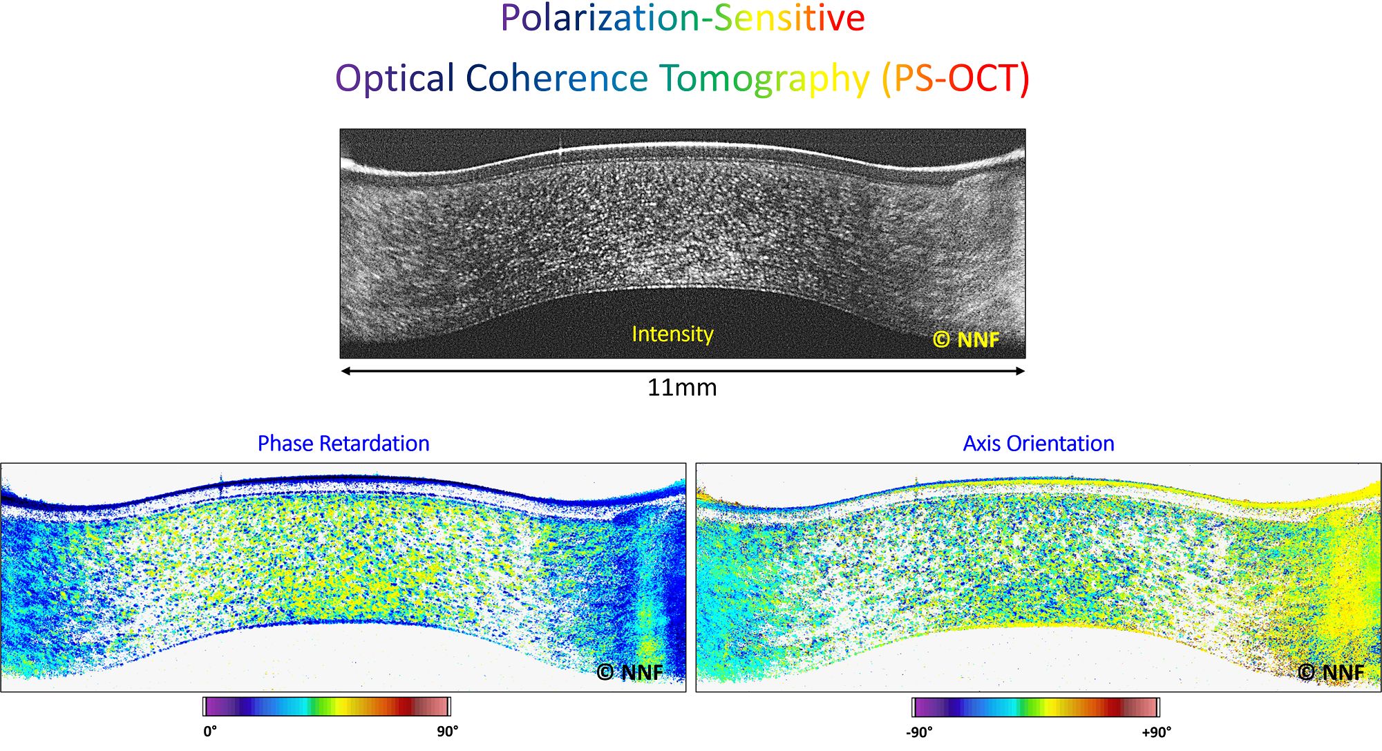
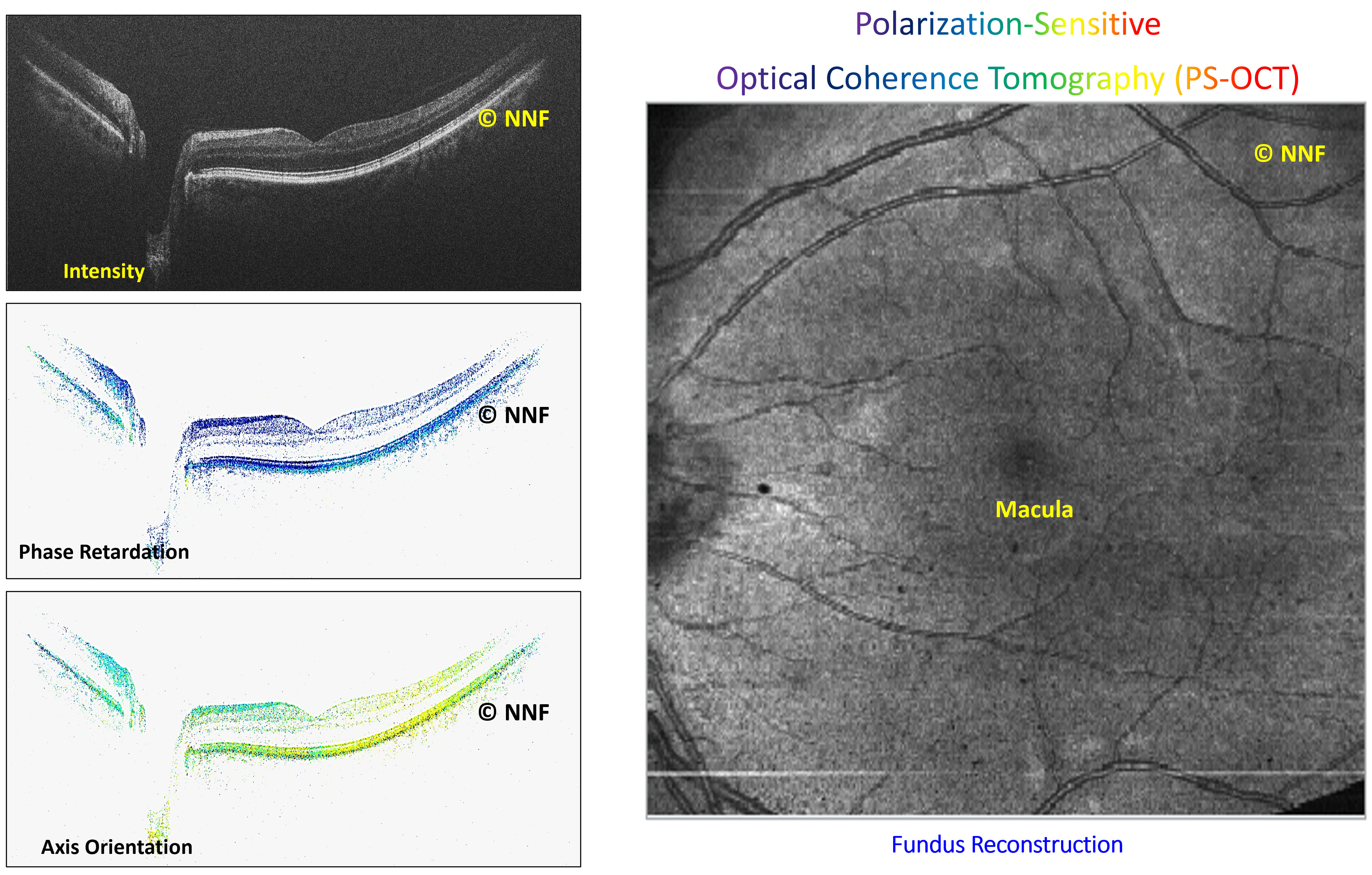
PS-OCT is a functional extension of conventional OCT, which assesses polarization-properties of fibrous tissue structures in cornea and retina, along with the usual intensity signals from OCT.

Our works with UHR PS-OCT mainly revolve around its clinical applications in ophthalmology.


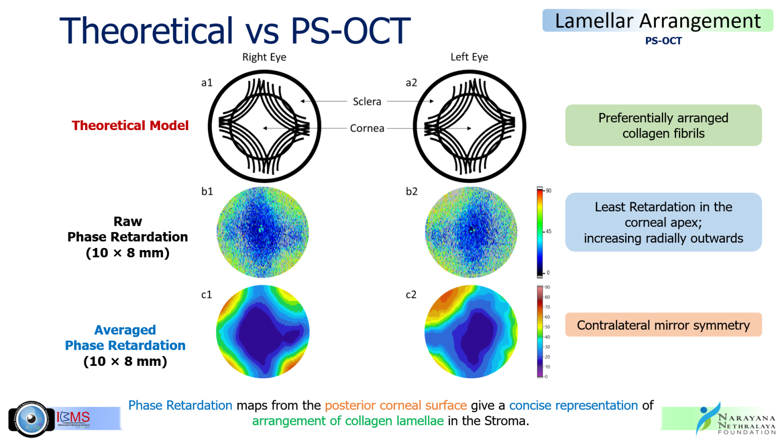
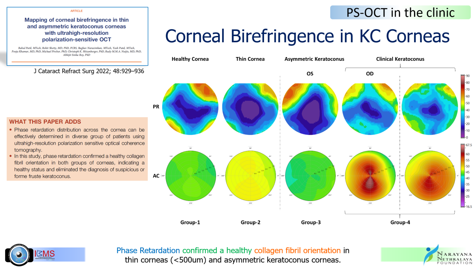
Phase Retardation (PR) is a measure of corneal birefringence. PR maps reconstructed from the volumetric data acquired from PS-OCT, are strikingly similar to the preferential arrangement of collagen lamellae described in ex vivo x-ray diffraction studies.

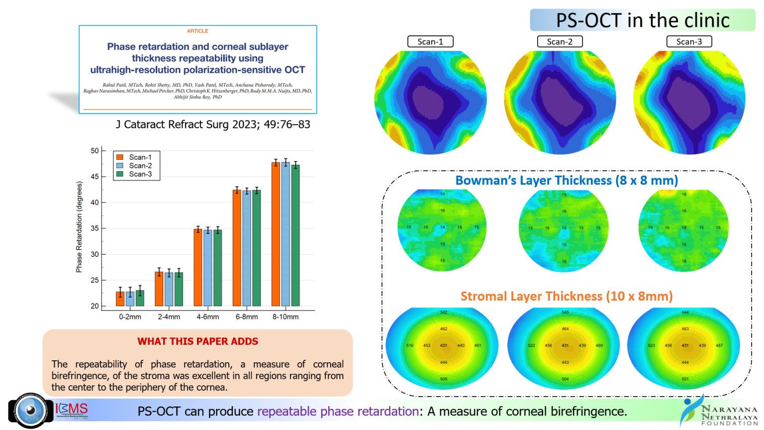
Our recent publication has shown the clinical stability of PS-OCT in generating repeatable and reliable phase retardation (PR) maps of healthy individuals with varying corneal thicknesses.


Owing to its ultra-high resolution capabilities, PS-OCT also provides detailed profiles of Bowman’s layer, stromal thickness, as well as epithelial and corneal thickness.

This helps the clinicians to know the “true” status of individual layers of the cornea and make an informed decision on further management.

The shape and transparency of our corneas are primarily dependent on the preferential lamellar arrangement and their corresponding fibril distributions. In degenerative diseases like KC, this preferential arrangement gets disturbed and worsens as the disease progresses. This alters the shape of the cornea and it continues to bulge outwards resulting in diminishing vision. 


Using UHR PS-OCT, one of our recent studies showed that even corneas with suspicious topographies and thin corneas showed good PR distribution indicating healthy preferential arrangement of collagen fibers, while the KC corneas showed significantly disrupted PR maps.


Research of this nature assists clinicians in making informed decisions regarding whether or not to proceed with eye surgery.

Through this website, we aim to provide you with a comprehensive overview of the latest advancements in ocular imaging, their applications in clinical practice, and their impact on patient care. Whether you are a healthcare professional, researcher, or simply interested in the field, we invite you to explore the articles, resources, and updates we have prepared for you.